As a Physiotherapist, I often use X-rays and other imaging techniques to help diagnose and understand the extent of arthritis in my patients’ knees. These images provide valuable insights into the condition of the joints and help guide treatment plans. In this blog post, we’ll take a closer look at X-rays and pictures of arthritic knees and discuss what we can learn from these images.
What is Arthritis of the Knee?
Arthritis in the knee, particularly osteoarthritis, is a condition where the cartilage that cushions the joints wears down over time. This leads to pain, swelling, and reduced mobility. Rheumatoid arthritis, another common type, is an autoimmune disease that causes inflammation of the joint lining. Both conditions can be identified and monitored through imaging techniques.
Understanding X-rays of Arthritic Knees
X-rays are a common diagnostic tool used to visualise the bones and joints. Here’s what we typically look for in X-rays of arthritic knees:
- Joint Space Narrowing: One of the primary signs of osteoarthritis is the narrowing of the space between the bones in the knee joint. This occurs as the cartilage wears away, reducing the cushion between the bones.
- Bone Spurs (Osteophytes): These are bony growths that develop along the edges of the bones. They are a common feature in osteoarthritic joints and can be seen as protrusions on the edges of the joint in X-rays.
- Subchondral Sclerosis: This refers to the hardening of the bone just below the cartilage surface. It appears as areas of increased bone density on the X-ray.
- Bone Erosions: In rheumatoid arthritis, X-rays might show areas where the bone has been eroded due to inflammation.
- Deformities and Misalignment: Severe arthritis can lead to changes in the shape and alignment of the knee joint. This might include bowing of the legs or other deformities visible on the X-ray.

Interpreting Pictures of Arthritic Knees
In addition to X-rays, photographs and MRI images can provide further insights into the condition of arthritic knees:
- Swelling and Inflammation: External pictures of arthritic knees often show visible swelling and inflammation around the joint. This is particularly common in rheumatoid arthritis.
- Redness and Warmth: Photographs can also capture redness and warmth around the knee joint, which are signs of active inflammation.
- MRI Images: Magnetic Resonance Imaging (MRI) provides a more detailed view of the soft tissues in the knee, including the cartilage, meniscus, and ligaments. MRI can detect early cartilage damage, meniscal tears, and synovitis (inflammation of the joint lining) that might not be visible on X-rays.

Case Studies: Interpreting an Arthritic Knee X-ray
Let’s take a look at an example X-ray of an arthritic knee and discuss what we see, The X-ray of the healthy knee above will help you compare.

- Joint Space Narrowing: Notice how the space between the femur (thigh bone) and tibia (shin bone) is significantly reduced compared to a healthy knee. This indicates cartilage loss.
- Bone Spurs: You can see small, pointed outgrowths along the edges of the joint, characteristic of osteophytes. Tibial spiking is similar
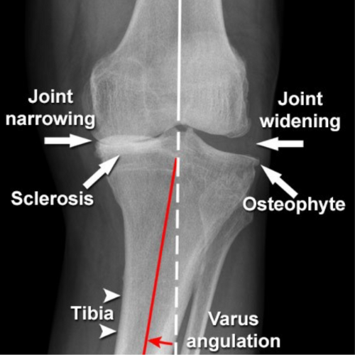
- Subchondral Sclerosis: There are areas of increased density below the joint surfaces, indicating subchondral sclerosis.
- Deformity: The alignment of the bones might appear altered, suggesting joint deformity due to advanced arthritis. Valgus (Knock Knees) or Varus (Bow legged)
Implications For Your Knee
Understanding the changes visible in X-rays and other images helps in formulating a treatment plan. However, x-rays can be very misleading. It’s good practice to treat the person and their symptoms rather than a scan or x-ray. The best way to look at this is by using real-life case studies.
WHICH ARE YOU? 1 or 2?
CASE STUDY 1
This elderly chap had one of the worst X-rays I had ever seen. But with no real pain to speak of he still lead a very active life. When I assessed his knee he had good strength and great alignment – so his knee although very arthritic was well-supported and relatively happy.
CASE STUDY 2
I see this regularly in clinic. People with very early arthritic changes but who are in agony. These people often have weak muscles around their knees and a posture which overloads their knees. Often these individuals have poor foot positions and lack strength and stability around their core and hips. Could this be you?
What does this mean? – HOPE
Just because you have an arthritic knee it does not mean you have to have pain and limited mobility.
YES – You hear me right.
I’m passionate about spreading this word because there is so much you can do to improve and offload your Arthritic knee.
Please don’t think it’s just a case of painkillers and waiting for surgery. It is not!
Join me on my FREE Masterclass learn how to manage & prevent a flare ups. We explore exercise and get started to your journey to better strength. Click to learn more and enrol.
Medical Treatment
There is a range of options available from full Knee Replacements, keyhole surgery, injections and more. I have a blog where we look in more detail at these options. Take a look if you are interested – Medical Treatments for Knee Arthritis: An Overview,
Conclusion
X-rays and images of arthritic knees provide critical insights into the extent and nature of the disease. By understanding what these images show, we can better diagnose the condition and tailor treatment plans to each patient’s needs. Hopefully, you now feel more empowered by better understanding your X-ray findings. Along with the knowledge that despite what your X-ray shows, you can still improve the health of your knees. If you have concerns about arthritis, consult with your healthcare provider for a thorough evaluation and appropriate imaging studies.
Take care, Helen
Helen Manders BSc (Hons) MCSP HCPC
Chartered Physiotherapist
Treating Arthritic Knees Since 2001